Cruciate ligament surgery
Treatment: Surgical Options
Cruciate ligament surgery is recommended for the treatment of rupture. The ligament can never regrow or repair, and unless surgery is performed the knee joint will remain unstable and degenerative arthritis will occur.
There are many surgical options available as part of the management of cranial cruciate ligament disease.
These varied techniques can be basically divided into two groups:
1) Lateral Suture Techniques in which a synthetic band is applied just outside the joint to mimic the action of ligament.
2) Bone Leveling Procedures which alter the geometry of the knee and allow it to function successfully without a ligament.
Below is a description of some of the surgical techniques that may be used to assist patients with cruciate ligament disease.
Based on your dog’s breed, size, weight and x-rays results, your vet will recommend which technique is most suitable for your dog.
1. Lateral Suture Technique
This technique involves the use of a prosthetic nylon ligament placed just outside the knee joint. The artificial ligament helps to prevent the forward motion of the tibia from underneath the femur when the patient walks ( like the cranial cruciate) Lateral Suture will ultimately break down but in the appropriate patients, it works well to stabilise the knee until the healing and scar tissue from the surgery provide enough support for the patient. For this reason, the Lateral Suture procedure is generally only performed in selected smaller dogs or cats.
Lateral Suture cruciate repair is performed at Southern Cross Vets by our own surgeons.
2. Bone leveling Procedures
For each of these different cuts are made in the top of lower bone (tibia) in order to make the cranial cruciate ligament unnecessary.
In each case, metal implants are placed into the leg and as with all orthopaedic procedures, a small number of patients may have complications as they recover. Complications are not at all common but may include fracture of the tibial tuberosity and implant infection post surgery.
Each of these procedures has a high success rate and most surgeons choose to select one of the procedures and develop great skill with that procedure, which results in the optimal outcome for the patient.
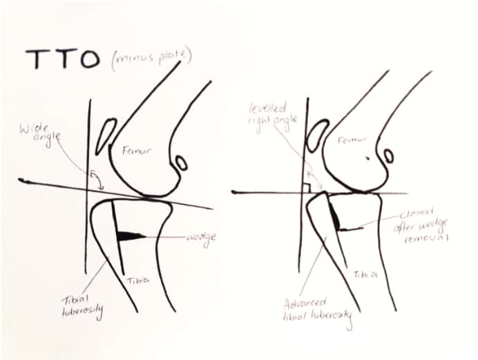
TTO
TTO stands for Triple Tibial Osteotomy.
TTO is an Australian-born innovation and is the newest way to repair a damaged cruciate ligament. It can be thought of as a hybrid of the TPLO and the TTA which we describe below) adding together the advantages of both procedures. The technique works by moving forward the top of the shin bone and realigning the top section of the shin bone.
During the surgery, the knee joint is opened and the delicate cartilages inside are examined. The remnants of the torn ligament may be removed, along with any damaged cartilage menisci present. An incision is then made vertically through the tibia (see diagram). A very small horizontal wedge of bone is also taken from within the tibia. The gap left by the wedge is gently closed by moving the edges of the bone together. This pushes out the tibial tuberosity and realigns the knee joint. A metal plate is screwed over the tibia to hold the changes in place and a bone graft is placed into the newly created gap in the bone. The surgeon will then suture closed the wound and take x-rays to confirm that the plate and bone lie in the correct position.
TTO is routinely performed at Southern Cross Vets by one of our own surgeons, Dr. Ed. as well as our Visiting Surgeon.
TPLO
This procedure that was developed in the United States, and Like a TTO, it modifies the knee joint to eliminate the need for a cranial ligament. In a TPLO a circular cut is made through the tibia. The top of the tibia is rotated down and secured in position with a metal plate to level the tibial plateau of the knee.
The TPLO is the procedure most often performed by American trained veterinarians.
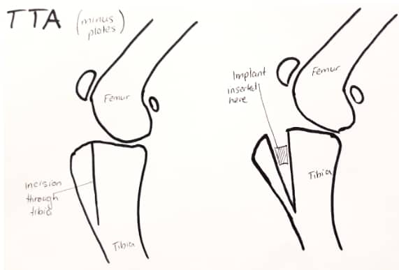
 TTA
TTA
TTA was developed in Europe and it stands for Tibial Tuberosity Advancement. Similar to TTO and TPLO, TTA aims to permanently alter the knee joint to stabilize it without a ligament. An incision is made through the tibia parallel to the tibial crest. A cage implant is inserted between the tibial tuberosity and the bulk of the tibia to advance the tibial tuberosity. Over time bone grows into the cage implant and the implant is incorporated as part of the bone. Removal of TTA implants can be difficult if complications develop bone has started to grow into the implanted mesh.
TTAs are not performed at Southern Cross and the reason for this are:
- The risk of developing a fracture mid surgery is too high
- The chance of a meniscus tear later in life is much higher that the other procedures. If a meniscus tear occurs, it almost always requires a second surgery.
- Dogs with a steep tibial slope do not benefit from this surgery
“The short story is that ALL the levelling procedures do a good job in reducing arthritis and immediately repairing the knee, the BEST levelling technique is the one that the surgeon has done most of. Our surgeons have collectively performed thousands of levelling procedures with an excellent success rate.”
Recovery
Most dogs will return to normal function by 16 weeks post surgery. For the first 6 weeks, strict crate confinement is required. This minimizes movement and allows the bone to heal correctly in its new position. You may be asked to perform physiotherapy exercise and massage the leg during this time. At the 6-8 week mark repeat x-rays are taken to check bone healing. Gradual return to exercise begins at 6-8 weeks after x-rays.
The aim of dog cruciate surgery recovery is to return to as close to 100% function as possible. Correct stabilization of the knee is important to minimize the development of arthritis. Unfortunately, in all cases of cruciate disease, some degree of arthritis is inevitable. Opting for the best surgery for your dog will likely be the difference between manageable arthritis in the distant future and unmanageable painful arthritis in the near future.
Things that we consider when choosing which surgery to perform:
- Pet’s weight – The higher the weight the more likely a levelling procedure will be better (almost all over 12kg is indicated)
- Shape of the shin bone – The more sloped the tibia, the more likely a levelling procedure is recommended regardless of weight
- Whether there is a meniscal injury – if the meniscus is injured, almost always we will recommend a levelling procedure to give the best pain relief (analgesia)